Case Examples

How SIJ cases can look completely different.

These case examples show how SIJ dysfunction can present through degeneration, prior lumbar fusion, failed pain procedures, hypermobility, and instability. The diagnosis often becomes clearer only when the history, examination, imaging, and response to injection are considered together.

Case 1.

Case 1 overview slide showing a 67-year-old patient with severe back pain

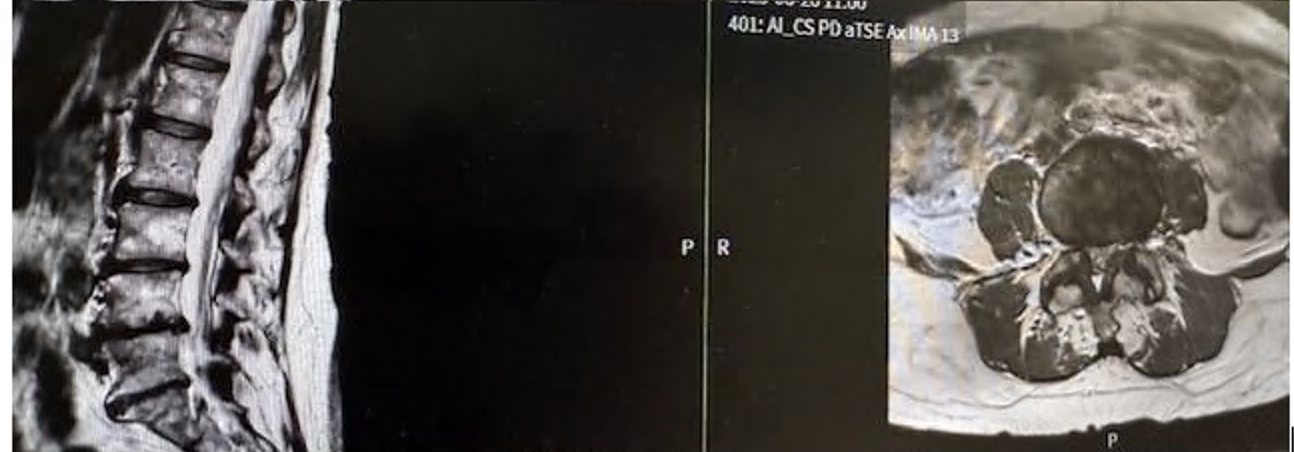
A 67-year-old patient from Melbourne presented with severe back pain rated 8/10 after failed physiotherapy, SIJ prolotherapy, and pain management.

  • SIJ injection gave only 24 hours of relief, but that response remained diagnostically important.
  • Beighton score: 7/9.
  • MRI showed severe discovertebral degeneration and facet arthritis.
Case 1 Outcome.

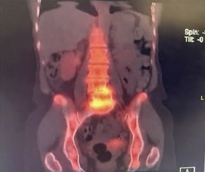
Case 1 follow-up imaging and treatment outcome after endoscopic radiofrequency ablation

Treatment progressed to endoscopic radiofrequency ablation and neurectomy from L3 to S3.

  • The patient reported an 85% improvement.
  • Follow-up imaging continued to show marked degenerative change at L4/5 and L5/S1.
  • This case highlights how SIJ pain can coexist with major lumbar degeneration.

Case 2.

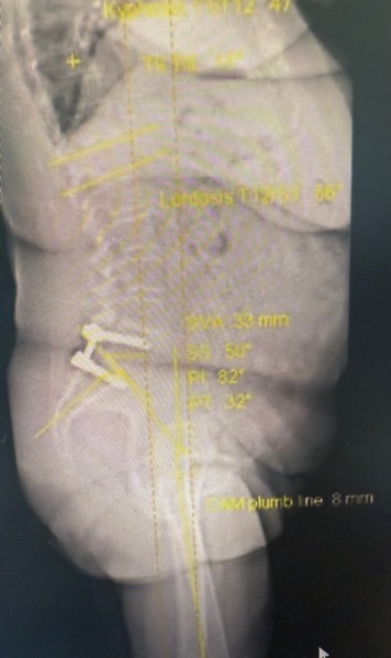
Standing radiograph from Case 2 showing prior lumbar fusion and spinopelvic alignment

A 75-year-old woman developed right-sided buttock, groin, and leg pain 33 years after an L5/S1 fusion.

  • MRI was limited by artifact.
  • CT did not show major compression.
  • She had excellent relief from an SIJ injection.
  • The patient underwent SIJ fusion with good relief of the pain.

Case 2 Imaging.

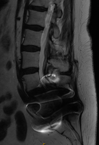
MRI for Case 2 demonstrating hardware artifact that complicates lumbar assessment

This case is a good example of a diagnostic dilemma after prior fusion, where imaging may not cleanly explain the pain pattern.

  • Prior spinal hardware can make MRI interpretation difficult.
  • CT may not reveal a compressive cause even when symptoms are significant.
  • The response to SIJ injection helped redirect the diagnostic pathway.

Case 4.

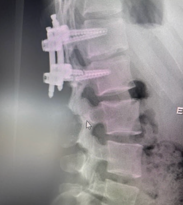
Radiograph from Case 4 showing hypermobility-related spinal issues

A 21-year-old gymnast had a broken screw at L1/2 from another surgeon, which was revised, but her original pain source was the sacroiliac joint.

  • She was treated with physiotherapy, SIJ injections, PRP injections, and radiofrequency ablations.
  • When everything failed, robotic sacroiliac joint fusion was performed with a good result.
  • Patients with hypermobility, including Ehlers-Danlos syndrome, can have severe ligamentous laxity and may fail non-surgical treatments.

Case 4 Treatment Path.

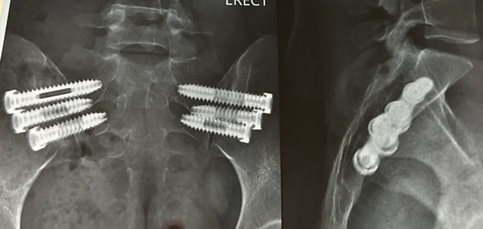
Post-treatment imaging for Case 4 showing sacroiliac fixation

The treatment journey included revision of the broken screw problem and multiple SIJ-focused non-surgical treatments before definitive progression.

  • L1/2 screw revision was performed, but this was not the original pain generator.
  • Non-surgical treatment included physiotherapy, SIJ injections, PRP injections, and radiofrequency ablations.
  • After failure of non-surgical treatment, robotic sacroiliac joint fusion gave a good result.

Case 5.

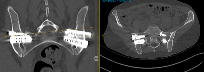
Case 5 CT image showing sacroiliac fixation in a patient with hypermobile EDS

A 38-year-old psychologist with hypermobile EDS reported severe back pain and instability over several years.

  • Prolotherapy gave only short-term improvement.
  • MRI of the lumbar spine appeared essentially normal.
  • She underwent SIJ fusion after failing all non-surgical treatments, with excellent results.
  • This case reinforces that major SIJ symptoms can exist despite relatively unremarkable lumbar imaging, especially in hypermobile patients.

What These Cases Show

Why SIJ diagnosis needs context.

Across these cases, the pain source was clarified by combining the clinical story with targeted testing instead of relying on one scan alone.

  • Older degenerative cases and younger hypermobile cases can both point strongly toward the SIJ.
  • Prior lumbar fusion can shift force and create diagnostic confusion later on.
  • Short-term relief after SIJ injection can still be clinically meaningful.
  • Normal or limited lumbar imaging does not rule out important SIJ dysfunction.